Marianas SDC

Super-Resolution Spinning Disk Confocal Microscopy System

Highlights

Spinning Disk Confocal

Yokogawa spinning disk confocals utilize a dual Nipkow disk with microlenses for the best optical sectioning and minimal pinhole crosstalk. This proven technology is the best solution for live cell imaging where optical sectioning and cell viability are both critically important.

CSU-X1

  • Highest speed imaging at up to 2000fps
  • Field of view for 7mm x 10mm detectors
  • 50µm pinhole disk with microlenses
  • Manual and motorized versions

CSU-W1

  • High speed imaging up to 200fps
  • Wide field of view 16mm x 17mm
  • 25µm and 50µm pinhole disks for lower and higher magnification objectives
  • Motorization including disk exchange, variable aperture, camera port selection and camera port magnification
  • Options for split-view imaging, NIR imaging, illumination field flattening and super-resolution imaging
CSU-X1 CSU-W1
Pinhole Diameter 50µm disk 25µm disk and 50µm disk
Number of Disks One One or two with motorized switching
Disk Bypass With 3i bypass Standard
Acquisition Speed 2000 FPS 200 FPS
Effective Field of View 10mm x 7mm 17mm x 16mm
Near IR Excitation Up to 640nm Up to 785nm

Uniformizer | Illumination Field Uniformity

For exceptionally even illumination across the entire field, Uniformizer conditions the gaussian beam from the illumination fiber optic to distribute light evenly across the field. Using a set of microlens arrays, Uniformizer flattens the field to as little as 1% variance and boosts overall intensity up to 50%.

Standard Montage

With Uniformizer

Prostate tissue section with nuclei in blue and vasculature in orange. Imaged with Marianas SDC and Uniformizer, 100x/1.46NA objective. Image shown is 650µm across montaged with 2 x 6 fields of view.

Super-Resolution by Optical Re-Assignment

CSU-W1 SoRa is an easy-to-use super-resolution microscopy solution utilizing a dual Nipkow disk assembly with microlenses on both the illuminating and pinhole disks. SoRa images have a 1.4x resolution improvement and deconvolved SoRa images have a 2x resolution improvement compared to standard spinning disk data. With a maximum speed of 200fps, low phototoxicity and no limitation on dyes or fluors, SoRa is ideal for super-resolution live cell imaging. SoRa is also available as an upgrade to existing CSU-W1 systems.

Specifications
Imaging Speed 200 fps
Wavelength Range 405nm to 640nm Excitation
420nm to 680nm emission
XY Resolution 150nm
120nm with deconvolution
Z Resolution 320nm
300nm with deconvolution
Field of View
63x Objective and 4x Relay Magnification
67µm x 63µm
Field of View
100x Objective and 2.8x Relay Magnification
61µm x 57µm

Standard

Standard Deconvolved

SoRa

SoRa Deconvolved

Argolight test slide (520nm emission) imaged with Plan-Apochromat 100x/1.46NA objective and 2.8x magnification. SoRa achieves a resolution of 150nm, improved to 120nm with deconvolution.
Imaging of microtubules in fixed bovine pulmonary artery endothelial cells. Azuma, T. and Kei, T. (2015) Super-resolution spinning-disk confocal microscopy using optical photon reassignment. Opt Express. Jun 1;23(11):15003-11. doi: 10.1364/OE.23.015003.

Photomanipulation

Ablate! Laser Ablation System

  • 355nm or 532nm pulsed laser
  • Fixed point or salvo-scanned variable region of interest
  • 2D or 3D regions
  • Diffraction limited spot

 
PHOTOABLATION
A Lattice lightsheet formed in fluorescent dye, viewed from the imaging objective.

Vector2 Scanning Photomanipulation

  • Photoactivation/FRAP
  • Galvo-scanned variable region of interest
  • 2D or 3D regions
  • Diffraction limited spot

 
PHOTOBLEACHING
Mammalian cell with membrane labeled in green before bleaching and FRAP curve in SlideBook.

Phasor Holographic Photomanipulation

  • Spatial light modulator-generated holography for optogenetics stimulation/FRAP/voltage imaging
  • Simultaneous 3D stimulation of multiple, separate regions
  • Visible and multiphoton stimulation without scanning
3D PHOTOSTIMULATION
3D illumination pattern (left) applied to a 3D specimen (right) to stimulate multiple regions simultaneously.

Technology

Adaptive Optics for Live Cell Microscopy

C-Shaper

Adaptive optics (AO) are used in astronomy to resolve images of distant celestial events through miles of light-scattering atmosphere. C-Shaper brings AO to live cell microscopy, improving images deep into specimens with seamless integration in Marianas systems. C-Shaper corrects classical optical aberrations due not only to imperfect lenses but due to unavoidable refractive index mismatches and unpredictable specimen environments. Correction can be done on-the-fly to address the changing optical environment that presents when imaging deep into a specimen.

Up to 18 Zernike modes can be addressed via an array of actuators in a deformable mirror. With the push of a button in SlideBook, aberrations are measured and corrected during image capture. Additionally, aberrations can be dynamically corrected as capture proceeds deeper into a specimen. The resulting improvements to the point spread function provide better resolving ability and increased signal to noise.

When imaging into a living cell with an oil objective, refractive index mismatch degrades the image quality. Addressing this via AO allows for instant correction of spherical aberration.

F-actin in fibroblasts imaged 50µm into water using a 100x/1.4NA oil immersion objective and CSU-W1 spinning disk confocal without AO (Left) and with AO correction (right).

Total Internal Reflection Fluorescence

Vector2 TIRF | Spinning X,Y TIRF System for Even Illumination

Vector2 TIRF is a flexible spinning TIRF (ring TIRF) addition to the Marianas platform. Vector2 TIRF uses galvo mirrors to spin an excitation laser around the periphery of the objective’s back focal plane. This produces an even evanescent wave with very low penetration depth, perfect for imaging live cell focal adhesions, membrane dynamics, and receptor function as well as in vitro experiments that require the thinnest optical section. The motorized focus of Vector2 TIRF allows for precise TIRF alignment and focus correction across all wavelengths and multiple objects.

Starting with a single point at the edge of the back aperture, Vector2 TIRF spins the beam around the periphery creating an evanescent field from all angles producing evenly illuminated images.

HILO Microscopy

Vector2 TIRF is also designed for highly inclined and laminated optical sheet (HILO) microscopy. HILO illumination (shown in orange) is achieved just before reaching the critical angle required for TIRF evanescent wave illumination (shown in blue). HILO offers high signal-to-noise with greater penetration for imaging beyond the membrane to the nucleus and through the cell body. Combined with LaserStack and a selection of high and low-powered lasers, Vector2 TIRF is a versatile single-molecule imaging platform in both TIRF and HILO modalities.

LightSheet

Marianas LightSheet

Marianas LightSheet incorporates a dual view single plane illumination microscope (SPIM) atop a fully automated research microscope. Light sheet illumination from sheets as thin as 3µm allow for long-term live cell imaging with essentially no photodamage. Dual view SPIM and image reconstruction in SlideBook produce data with isotropic 3D resolution.

TILT Stage Top LightSheet

TILT is a single objective lightsheet offering convenience and flexibility across a variety of magnifications and numerical apertures. Easily incorporated with Marianas, TILT produces light sheet illumination of less than 5µm sheet thickness nearly normal to the imaging objective optical axis.

Frequency Domain FLIM

Fluorescence lifetime imaging microscopy (FLIM) for Marianas measures fluorescence lifetimes via frequency modulation, creating data orders of magnitude faster than time domain FLIM. Frequency domain FLIM modulates both illumination and detection signals for artifact-free FRET and micro-environment sensor imaging in live samples. Lifetime images can be acquired in less than one second and close to single-molecule detection is possible.

Fluorescence lifetimes can be used to measure protein proximity (FRET), aggregation, relative concentration of different molecules, separation of different markers with spectral overlap, ion concentration, and for the removal of autofluorescence. SlideBook software allows FLIM imaging to be fully integrated and automated, combinable with widefield fluorescence, TIRF, FRAP, spinning disk confocal and multiphoton imaging. Through intuitive polar graph (vector) graphic analysis, SlideBook can easily visualize a variety of fluorescent substances, FRET efficiency and mixture analysis.

FLIM
c1-6ab Hela cells dual expressing donor and acceptor FRET pair. 488nm illumination. Elenora Balloi, Peter March Lab, Welcome Trust, Manchester UK.

Plant Microscopy

Marianas can be outfitted with a vertical stage and a spinning disk confocal for high resolution 4D imaging of developmental biology in Arabidopsis and other plants. A stage designed for plant chambers and a right-angle relay from the microscope allow long-term study of plant physiology in their native growth orientation.

PLANT BIOLOGY
Actin filaments, labeled with mNeonGreen-fABD, in epidermal root cells. Courtesy of Dr. Felix Ruhnow and Isabella Østerlund, Dr. Staffan Persson group at University of Copenhagen.

Nano Injection

Single Cellome Unit SU10

Micromanipulators can be added seamlessly into Marianas experiments. The SU10 is a next generation minimally invasive intracellular nano-injector glass pipette, with a tip size of under 100nm. It can be programmed for automated cell surface detection, penetration and delivery

Features

Slide

Up to four fiber outputs
Millisecond path switching

Environmental Control

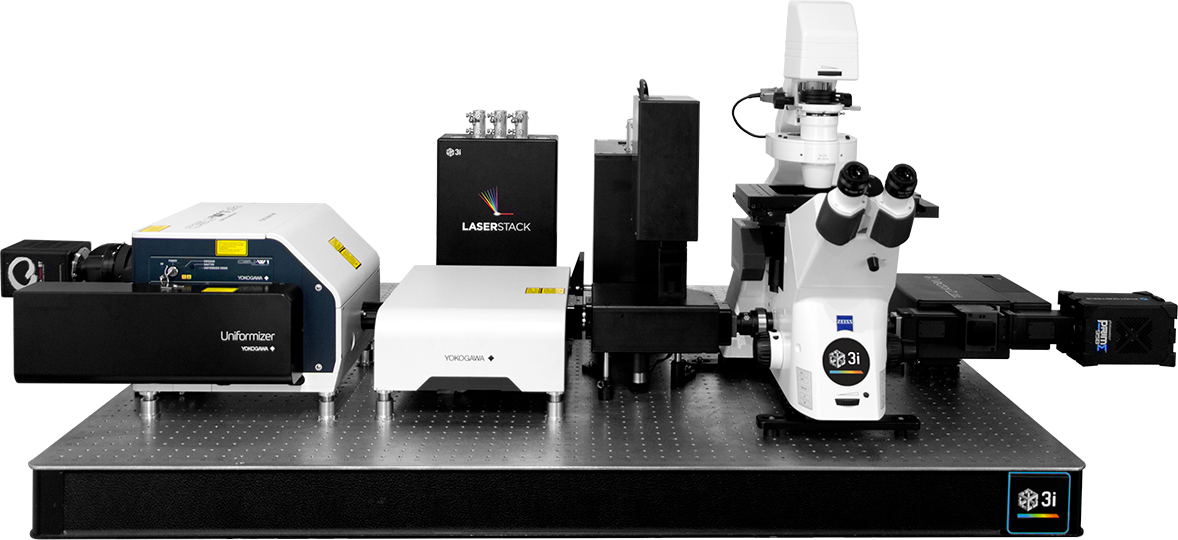
Fully Automated Microscope

Computer generated holography
1-photon and 2-photon

Stage top and cage incubators
Temperature, gas and humidity

Motorized objective, condenser, path selection
Autofocus (Definite Focus 3)
PSF-optimized objectives

Laser ablation system
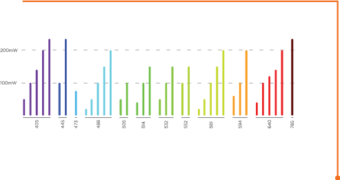
355nm and 532nm

Spinning total internal reflection fluorescence

Modular high-speed X/Y scanner

Millisecond timing and trigger
Control of multiple devices

Modular laser combiner
Up to eight lasers

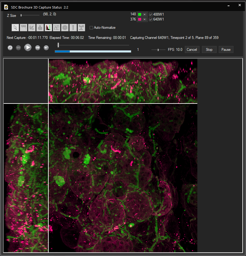
Live cell 3D confocal imaging
Super-resolution dual microlens disk

Path selection up to four ports
Sub-millisecond switch time

Dynamic correction of spherical aberration with depth

SlideBook Software for Acquisition and Analysis

SlideBook software supports research microscopy through the entire experimental process. By managing everything from instrument control to image processing and data analysis, SlideBook allows scientists to focus on investigation rather than instrumentation. SlideBook controls hundreds of instruments in and around the microscope from dozens of manufacturers enabling researchers to integrate their preferred components and upgrade to the latest devices once available.

Slide

User-Selectable App Appearance

Select a color scheme from dozens of options
Switch on-the-fly from dark to light themes

SlideBook Open File Format

Directory-based open file format for big data and high performance computing applications

Volume Rendering

3D and 4D volume view visualization tools support a user- specified bounding box and a storyboard interface where multiple perspectives can be assembled into a single movie

NVIDIA CUDA GPU Acceleration

GPU acceleration of computationally-intensive operations such as deconvolution

System Capture Consoles

Consoles are a single easy-to-use window featuring all frequent controls and status displays

3D Capture Status

Multiwell and Montage

Volumetric projection during 4D capture supported across all instruments

Streamlined multiwell interface
Montaging with a variety of methods

Capabilities

Capture

Control hundreds of devices including microscopes, stages, lasers, wheels, piezos, scanners, shutters and much more.

View

Visualize data through any numbers of portals, from single images to z-stacks, time lapse, color channels and 4D views.

Analyze

Analyze images and extract statistical data via a wide variety of algorithms while maintaining original data integrity.

Scripting

Macro scripting for capture and analysis enhances the flexibility and power available to users.

Communicate

Present and export data easily as 16-bit TIFFs, 3D movies, graphs or spreadsheets. Data is directly portable to MATLAB and Excel and adheres to Open Microscopy Environment (OME) standards.

Partners

MATLAB

Through hierarchical and conditional capture, user-supplied MATLAB programs can control experimental workflows.

Microvolution

Microvolution software delivers nearly instantaneous deconvolution by combining intelligent software programming with the power of a GPU.

Aivia

Aivia is an innovative and complete 2D-to-5D image visualization, analysis and interpretation platform with artificial intelligence-guided image analysis.

Dell

The latest high-power computer workstations control all microscope hardware and enable high-speed processing, segmentation and volume rendering of terabyte (TB) datasets.

Powerful Computer Workstation

3i provides high-power computer workstations to control all microscope hardware necessary for acquisition as well as enable processing, segmentation and volume rendering of terabyte (TB) datasets without additional computer resources.  Solid state drives in RAID configurations provide high-speed storage for capture, while traditional high-capacity hard drives provide longer-term storage of datasets. NVIDIA Quadro GPUs work in parallel with the latest Intel Xeon processors for fast stitching, processing and rendering of captured data.

Petabyte Data Storage

3i offers DDN® unified storage systems to allow direct acquisition and analysis, without time-consuming file transfers, at volumes ranging from 500TB to over 2PB. DDN storage systems are an ideal choice for labs and facilities looking to optimize acquisition workflows and/or incorporate data analysis pipelines.

Marianas SDC Data Sets

PLANT BIOLOGY
Actin (magenta) and microtubule cytoskeleton (cyan) in Arabidopsis thaliana cotyledons. Courtesy of Isabella Østerlund and Dr. Felix Ruhnow, Dr. Staffan Persson group at University of Copenhagen.
Ex Vivo
Murine hair follicles. Courtesy of Dr. Anthony Peng, Massachusetts Institute of Technology.
Intracellular Imaging
Macrophage labeled for nucleus (blue), microtubules (purple), and actin (orange).
Cancer Research
Prostate tissue section labeled for nuclei (blue) and blood vessels (orange).
Reproductive Medicine
Lipid docks in lactating mammary epithelial cells. 20x montage. Courtesy of Dr. Jenifer Monks, CU Anschutz Medical Campus.
 
Developmental Biology
Drosophila pupa wing disc expressing E-cadherin GFP.
Developmental Biology
Zebrafish tail labeled for actin.
CELL BIOLOGY
Retinal pigment epithelium cells expressing GFP-vimentin (green) and RFP-tubulin (blue). Courtesy of Dr. Doncic Lab, University of Texas Southwestern Medical Center.

Additional Resources

Find out more about Marianas SDC


    SALES

    Email: sales@intelligent-imaging.com

    Phone: +1 (303)-607-9429 x1

    SUPPORT

    Email: support@intelligent-imaging.com

    Phone: +1 (303)-607-9429 x2