


Up to four fiber outputs
Millisecond path switching
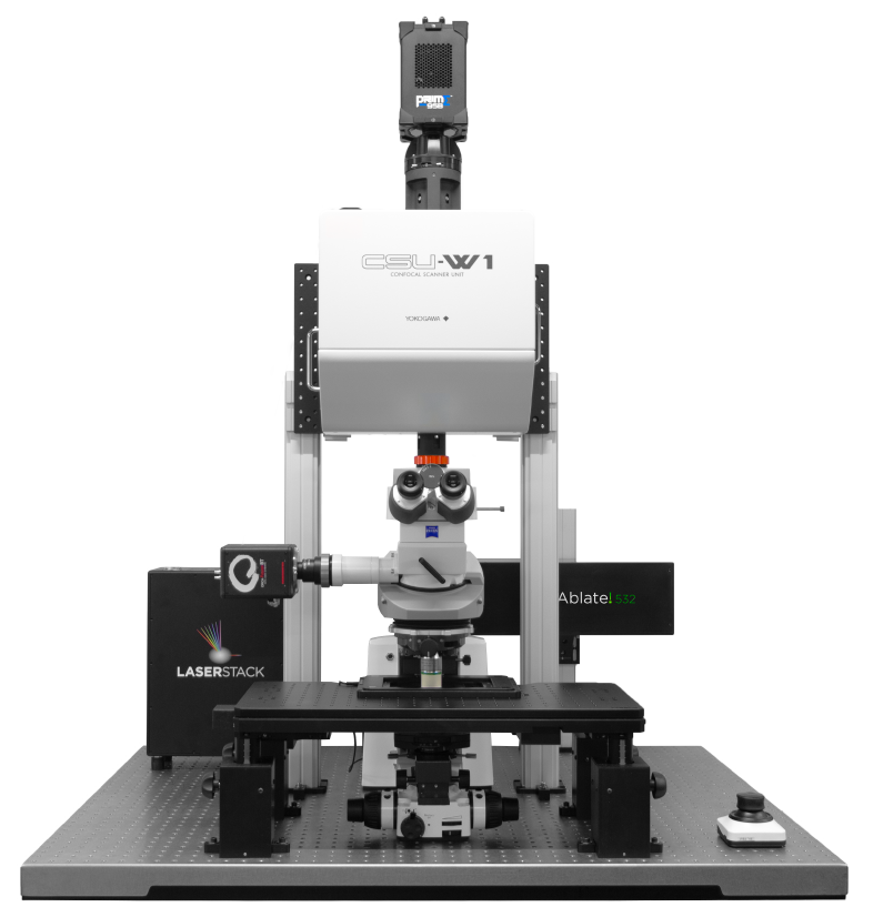
Fully Automated Microscope
Platform Stage
Telecorr

Computer generated holography
1-photon and 2-photon
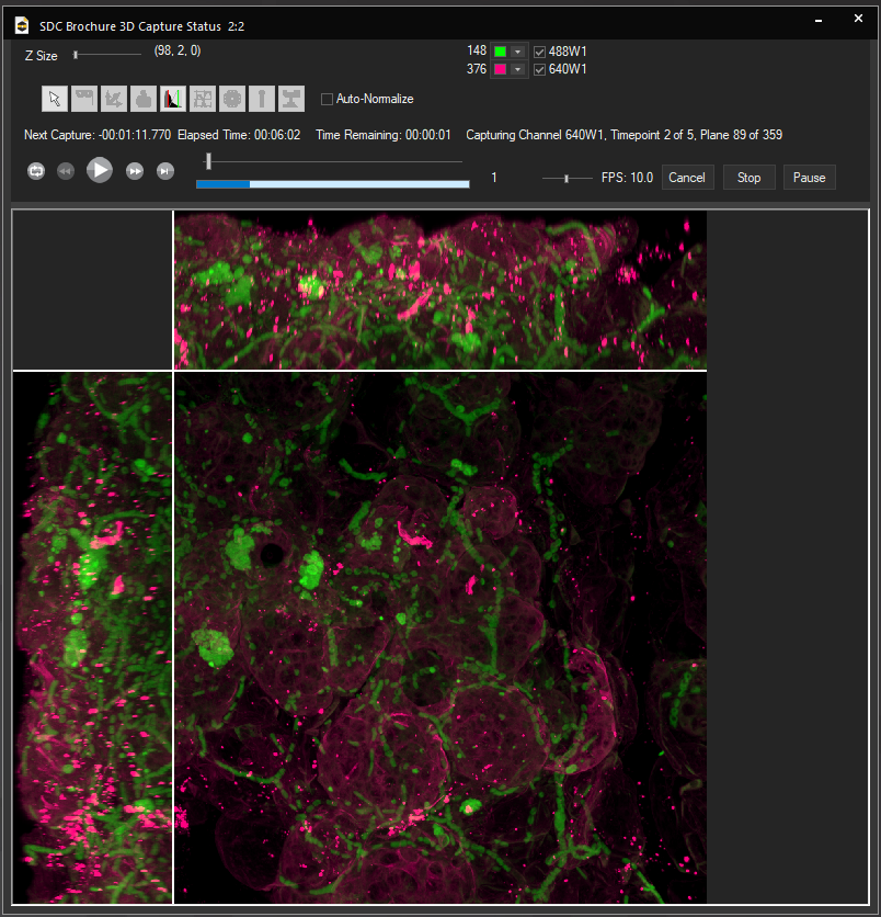
Intravital 3D confocal imaging
Super-resolution dual microlens disk
Motorized z-drive (with optional fast piezo)
Long working distance water dipping objectives
Large, easily accessible sample space
355nm and 532nm pulsed laser system
Fixed point or galvo-scanned for fast targeting
Accommodates intravital imaging trays and life support systems for long-term animal imaging
Modular high-speed X/Y scanner for accurate, diffraction-limited photomanipulation events


Millisecond timing and trigger
Control of multiple devices
Modular laser combiner
Up to eight lasers

Surgical Trays
Custom-designed surgical trays for presentation of exteriorised tissue and for stabilization of tissue presentation using cranial windows

Spinning Disk Confocal
Focus extender for correct spinning disk confocal excitation