Lattice LightSheet

A Microscope for High-Resolution, Fast, and Gentle 3D Live Cell Imaging

Break the Diffraction Limit with SIM

Single-Molecule Imaging

Photomanipulation

A Microscope for High-Resolution, Fast, and Gentle 3D Live Cell Imaging

Lattice LightSheet (LLS) is a cutting-edge light sheet microscope built for cell biologists. The system generates an optical lattice to create an ultra-thin light sheet to image biological samples over long periods of time and with very fine resolution. This allows for 4D live cell imaging, where experiments limited to seconds or minutes on other imaging platforms can be extended to hours or even days. The combination of high spatiotemporal resolution, imaging speed and sensitivity make Lattice LightSheet the ultimate imaging tool for a new era of live cell microscopy.

Slide

Beam Steering

Fast X and Z galvanometers for rapid volumetric captures
Structured illumination for super-resolution imaging

Sensitive Cameras

Environmental Control

95% QE low read noise sCMOS detectors
Fast, sensitive spectral separation
Up to 2 cameras

Sample chamber and objective heating
Digital temperature, gas, and humidity control

Motorized Light Sheet Selection

Upright Microscope Configuration

Digital Light Sheet Formation

Standard glass sample mounting for cells and tissue
Custom sample mounting on other materials
Highest resolution, water-dipping objective pair
Ideal for live cells, organoids, tissue, and embryos

Beam structuring via spatial light modulator (SLM)
Square, Hexagonal, Bessel & other patterns

Adjustable light sheet length and thickness for a variety of samples, without realignment

Widefield Imaging Path

Inverted widefield path for easy sample finding
Brightfield LED & epi-fluorescence LED options

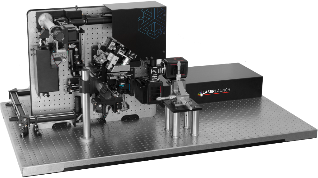
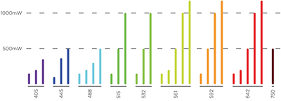
LaserLaunch Modular Laser Combiner

Highlights

Super-Resolution Imaging | Break the Diffraction Limit with SIM

Standard Sheet Scan

The lightsheet is rapidly dithered along the X axis and one image is captured per Z plane.

Deconvolution

Quantitative image restoration (after standard sheet scan) via constrained iterative deconvolution in SlideBook.

Structured Illumination Microscopy (SIM):

The light sheet is moved in five discrete phase steps along the X axis. Five raw images are collected that are reconstructed to produce an image that is beyond the diffraction limit of the detection objective by a factor of ~1.4x.

SlideBook acquires images with a stepped pattern. The step size is determined by the spacing of each pattern (wavelength and annular mask position). This is repeated throughout the entire Z-stack to acquire a full volume. SlideBook easily processes the raw SIM data into a super-resolved final image.

Standard

Standard Deconvolved

SIM

Mammalian cells expressing GFP-tagged lysosomes.

Photomanipulation

Photomanipulation is a powerful tool for studying membrane dynamics, receptor and vesicle movement and photo conversion of specific molecules. Addition of the Vector2 X,Y galvo scanner makes complicated bleaching, FRAP, uncaging and photoconversion experiments easy. Compared to a spinning disk or point scanning confocal, photomanipulation experiments on a lattice can be done more rapidly and with less inherent bleaching. Vector2 can be added to new or existing Lattice LightSheet systems.

 
photomanipulation
Bleaching of a cell membrane via Vector2. Targeting of specific regions of interest is highlighted by the flashes of excitation light in a line across the sample, repeated over a few time points to gently bleach out the target portion of the sample. Bleaching is possible with any laser present on the system.

 
frap
Fluorescence Recovery After Photobleaching (FRAP) can be used to study kinetics of membrane diffusion after a targeted region has been bleached of fluorescent signal. Over time the fluorescence in the targeted area recovers based on the rate of diffusion.

Single Molecule Localization Microscopy

Cell biologists and biophysicists are increasingly interested in tracking single molecules of interest instead of looking at groups of proteins as a unit. Single molecule localization microscopy (SMLM) offers researchers the ability to observe individual fluorophores on a single molecule of interest and observe how each behaves. Typically, techniques like TIRF have been required for SMLM but Lattice LightSheet offers the following capabilities to increase overall signal-to-noise and facilitate SMLM imaging in 3D:

  • High-powered lasers (up to 2W)
  • The most sensitive sCMOS cameras with low read noise and 95% QE
  • 100+ fps acquisition for rapid single plane imaging or acquiring volumes per second
  • Photoswitching and photoconversion through Vector2

Intracellular to Extracellular to Multicellular Imaging

Motorized Light Sheet Selection

With 17 different light sheet lengths and thicknesses to pick from, it is easy to find the ideal Lattice for your sample.

  • 15-20 µm sheets for intracellular events
  • 30-40µm sheets for small organoids and cell-cell interactions
  • 50-75µm sheets for large organoids and portions of embryos or larger organisms

Using the “Mask” mode allows for changing of the light sheet while the sample remains in focus, allowing users to evaluate different light sheet parameters in real time. SIM is possible with any of the sheets, opening the door for fast super-resolution imaging of an assortment of sample types.

 
Mitochondria
Mitochondrial fusion and fission is a rapid cellular process that is also indicative of cellular damage from phototoxicity. The gentle nature of the Lattice LightSheet slows for unprecedented, volumetric imaging of this dynamic process without photodamage. Mitotracker Red in RPE1 cell.

 
Immunology
Visualizing cell-cell interactions is commonly displayed using a volume rendering. Here a slice/ortho view across X, Y, and Z allows for detailed examination of each cell’s membrane due to the high axial resolution offered by the Lattice LightSheet. Jurkat cell (red) interacting with HeLa cell (green).

SLM Pattern Generation

A key feature of the Lattice LightSheet is the spatial light modulator (SLM), which generates interfering multi-Bessel patterns.

For most imaging, the traditional “square” lattice pattern offers a nice blend of low light dose, optical sectioning and high axial resolution. Innovations into new patterns is an ongoing topic and has created other designs know as “hexagonal” and “hex-rect patterns. These designs allow for fine tuning of axial resolution, SIM performance and deconvolution.

These non-traditional patterns can be easily used on the Lattice LightSheet by updating the pattern file loaded to the SLM. Since the patterns can be quickly exchanged, users can evaluate their qualitative performance in real time.

 
MULTICELLULAR
Medium-sized samples are easy to accommodate via longer light sheets, all controlled digitally. Drosophila primary neurons with Jupiter (orange) and CD8 (blue).

 
Plant Biology
Large samples, even with light scattering cell walls, can still be visualized with the Lattice LightSheet. Taking advantage of the longer 75 µm long sheet, a portion of a root tip expressing GFP tubulin is imaged in high resolution.

Technology

First developed by Nobel Laureate Dr. Eric Betzig, the 3i Lattice LightSheet microscope is capable of imaging biological systems spanning four orders of magnitude in space and time. The system generates an optical lattice to create an ultra-thin light sheet to image biological samples over long periods of time and with very fine resolution. This allows for 4D living cell imaging, where experiments limited to seconds or minutes on other imaging platforms can be extended to hours or even days. The combination of high spatiotemporal resolution, imaging speed and sensitivity make Lattice LightSheet the ultimate imaging tool for a new era of living cell microscopy.

Imaging Through a Lattice LightSheet

The Lattice LightSheet is a system designed to push the spatial and temporal resolution limits in live cell imaging. 3i has evolved the original design from Dr. Eric Betzig with the goal of making the system more user friendly.

1. Cylindrical lenses stretch and collimate the beam to form a sheet projected onto a spatial light modulator (SLM).
2. The SLM generates an optical lattice of Bessel beams.
3. The annular mask acts as a zero-order filter, removing artifacts and lengthening the sheet.
4. Galvos dither the sheet in X and sweep the sheet in Z.
5. A lattice sheet is formed over the sample space.
6. Emitted fluorescence is detected on a high QE, low read-noise, sCMOS camera.


Interfering Bessel Beams | Evolving the Gaussian

Light sheet microscopy involves illuminating a specimen orthogonal to the plane of detection. The beam is then quickly scanned or passed through a cylindrical lens to create a sheet of light. This Gaussian light sheet offers inherent optical sectioning and more chances for the excitation light to create fluorescent signal as it travels through the sample.

Using more exotic beam shaping, specifically Bessel and Multi-Bessel (Lattice) light sheets, the axial resolution and optical sectioning of the system can be drastically improved with minimal excitation power. The 3i Lattice LightSheet makes implementing these improved light sheets easy for the user, allowing scientists to focus on scientific exploration. The thin lattice light sheets are ideal for cell biologists pushing their experiments faster and longer than previously possible.

Gaussian

Gaussian beams are simple to create but are very short when thin (length is proportional to thickness).

Bessel

Bessel beams have great axial resolution and are uniformly thin but have a large amount of out of focus light, leading to a higher light dose on the sample.

Lattice

Interfering Bessel beams create an optical lattice that is still uniformly thin with great axial resolution but without the large amount of out of focus light. Lattice beams can also be used for structured illumination.

Specialized Objective Pair

The Lattice LightSheet’s two objectives are specifically selected and designed to complement the ultra-thin light sheet and offer high-resolution imaging. Using a light sheet as thin as 400nm further improves the axial resolution.

Illumination
Custom 0.71NA long working distance water immersion objective for lightsheet illumination, mechanically and optically matched to the imaging objective.

Imaging
High-resolution 1.1NA water immersion objective with depth of field matched to lightsheet thickness for excellent optical sectioning.

SlideBook Software for Acquisition and Analysis

SlideBook manages every step in Lattice LightSheet imaging. A comprehensive control module guides users through light sheet and beam selection, the collection of 3D and 4D captures, image processing, volume rendering and finally movie making with story-board support. SlideBook is GPU optimized and readily handles the creation and processing of 3D/4D datasets over 1TB, making them ready for analysis and rendering. SlideBook SLD and SLDY files can be accessed via applications supporting BioFormats OME and Python, allowing seamless collaboration in any workflow.

Slide

User-Selectable App Appearance

Select a color scheme from dozens of options
Switch on-the-fly from dark to light themes

SlideBook Open File Format

Directory-based open file format for big data and high performance computing applications

Volume Rendering

3D and 4D volume view visualization tools support a user- specified bounding box and a storyboard interface where multiple perspectives can be assembled into a single movie

NVIDIA CUDA GPU Acceleration

GPU acceleration of computationally-intensive operations such as deconvolution

Image Processing

Deskewing and rotation of data are built into SlideBook’s workflow

Multiposition Capture

System Capture Console

Intuitive controls for selecting multiple XYZ positions for automated, sequential capture, ideal for overnight experiments of dozens of locations

The LLS user interface is a single, easy-to-use window featuring all frequent controls and alignment tools from laser selection, system calibration, and dithered/SIM capture mode

Capabilities

Capture

Control hundreds of devices including microscopes, stages, lasers, wheels, piezos, scanners, shutters and much more.

View

Visualize data through any numbers of portals, from single images to z-stacks, time lapse, color channels and 4D views.

Analyze

Analyze images and extract statistical data via a wide variety of algorithms while maintaining original data integrity.

Scripting

Macro scripting for capture and analysis enhances the flexibility and power available to users.

Communicate

Present and export data easily as 16-bit TIFFs, 3D movies, graphs or spreadsheets. Data is directly portable to MATLAB and Excel and adheres to Open Microscopy Environment (OME) standards.

Partners

MATLAB

Through hierarchical and conditional capture, user-supplied MATLAB programs can control experimental workflows.

Microvolution

Microvolution software delivers nearly instantaneous deconvolution by combining intelligent software programming with the power of a GPU.

Aivia

Aivia is an innovative and complete 2D-to-5D image visualization, analysis and interpretation platform with artificial intelligence-guided image analysis.

Dell

The latest high-power computer workstations control all microscope hardware and enable high-speed processing, segmentation and volume rendering of terabyte (TB) datasets.

Powerful Computer Workstation

3i provides high-power computer workstations to control all microscope hardware necessary for acquisition as well as enable processing, segmentation and volume rendering of terabyte (TB) datasets without additional computer resources.  Solid state drives in RAID configurations provide high-speed storage for capture, while traditional high-capacity hard drives provide longer-term storage of datasets. NVIDIA Quadro GPUs work in parallel with the latest Intel Xeon processors for fast stitching, processing and rendering of captured data.

Petabyte Data Storage

3i offers DDN® unified storage systems to allow direct acquisition and analysis, without time-consuming file transfers, at volumes ranging from 500TB to over 2PB. DDN storage systems are an ideal choice for labs and facilities looking to optimize acquisition workflows and/or incorporate data analysis pipelines.

Lattice LightSheet Data Sets

 
Mitosis
The near isotropic resolution of the Lattice LightSheet allows for incredible videos of mitotic cells in 4 dimensions. Mammalian cells expressing H2B-RFP. Courtesy of Dr. Andrew McAnish, University of Warwick.
 
Membrane Dynamics
Actin ruffles on a large macrophage imaged at 1 volume/second to capture rapid membrane dynamics over a large cell. Macrophage with LifeAct GFP. Courtesy of Dr. James Springfield, University of Queensland.
 
Neuroscience
Imaging the process of neuronal axon lengthening requires a gentle excitation source and rapid volumetric imaging. Sample was imaged at >1 volume/second to capture rapid microtubule dynamics in the growth cone of a primary rat neuron.
 
Hematology
Platelet generation is typically a difficult process to capture using fluorescent microscopy – the megakaryocytes won’t produce platelets if they are excited with too much laser light. Here, the Lattice LightSheet is gentle enough to image this unique developmental process at high resolution. Courtesy of Dr. Watson, University of Birmingham.
 
Model Organism
Dictyostelium are a “slime mold” that tend to run away from exciation light before they begin to photobleach. The Lattice LightSheet is gentle and fast enough to image dictyostelium at over a volume/second without the cells migrating away. This allows for unprecedented imaging of this model organism. Dictyostelium expressing LifeActGFP. Courtesy of Dr. Till Bretschneider, University of Warwick.
 
MEMBRANE DYNAMICS
HeLa cells transfected with GFP-Lifeact (488nm) to visualize actin dynamics and stained with DiL (561nm red)(Thermo) to visualize membrane dynamics. Courtesy of James Springfield, Institute for Molecular Bioscience (IMB) at the University of Queensland
 
cytoskeleton
Microtubules are the ever changing cytoskeleton present in the cell. Here, fast volumetric imaging of the microtubule network displays distinct differences in the cytoskeletal dynamics through out the cell. MCF-7 cells transfected with GFP-EMTB to visualize microtubule dynamics. Courtesy of Dr. Gordon L. Hager and David A. Garcia at the National Cancer Institute.

Specifications

LIGHT SHEET THICKNESS

0.4µm at 50µm length

DETECTION OPTICS

1.1NA water objective, 2.0mm WD, 62.5x total magnification

ILLUMINATION OPTICS

0.71NA water objective, 3.7mm WD

LASER OPTIONS

Up to 6 laser lines, wavelengths from 405 – 670 nm, powers from 100 – 2000 mW (depending on wavelength)

STANDARD CAMERA

Hamamatsu ORCA-Fusion BT sCMOS Camera

CAMERA OPTIONS

Single sCMOS, Dual sCMOS direct 1x projection, Dual EMCCD relayed 2.5x projection

SAMPLE CHAMBER

Medical grade stainless steel with TEC temperature control and perfusion capabilities. Optional – complete environmental control system with active temperature, humidity, and CO2 control via Okolab.

SPECIMEN MOUNTING

Standard, horizontally-oriented 5mm round coverslip

ACQUISITION COMPUTER

Dual 16-Core Xeon Gold 2.9GHz processors, 128GB RAM, 8GB NVIDIA Quadro RTX4000 workstation graphics card, 512GB OS SSD, 8TB Fast Acquisition Drive, and 20TB additional storage

ANALYSIS COMPUTER

Dual 26-Core Xeon Gold 2.1GHz processors, 256GB RAM, 24GB NVIDIA Quadro RTX6000 workstation graphics card, 512GB OS SSD, 8TB Fast Acquisition Drive, and 20TB additional storage

STORAGE SOLUTIONS

DDN® unified storage systems for direct full-speed acquisition and analysis starting at 500TB. DDN systems utilize a BioScaler GPFS file system and are easily expandable to multiple petabytes

Publication Spotlight

CENP-F Stabilizes Kinetochore-Microtubule Attachments and Limits Dynein Stripping of Corona Cargoes.

Auckland, Philip, Emanuele Roscioli, Helena Louise Elvidge Coker, and Andrew D. McAinsh. “CENP-F Stabilizes Kinetochore-Microtubule Attachments and Limits Dynein Stripping of Corona Cargoes.” Journal of Cell Biology 219, no. 5 (May 4, 2020).

https://doi.org/10.1083/jcb.201905018

Insight from the Maximal Activation of the Signal Transduction Excitable Network in Dictyostelium Discoideum.

Edwards, Marc, Huaqing Cai, Bedri Abubaker-Sharif, Yu Long, Thomas J. Lampert, and Peter N. Devreotes. “Insight from the Maximal Activation of the Signal Transduction Excitable Network in Dictyostelium Discoideum.” Proceedings of the National Academy of Sciences of the United States of America 115, no. 16 (17 2018): E3722–30.

https://doi.org/10.1073/pnas.1710480115

Oxygen Tension and the VHL-Hif1α Pathway Determine Onset of Neuronal Polarization and Cerebellar Germinal Zone Exit.

Kullmann, Jan A., Niraj Trivedi, Danielle Howell, Christophe Laumonnerie, Vien Nguyen, Shalini S. Banerjee, Daniel R. Stabley, Abbas Shirinifard, David H. Rowitch, and David J. Solecki. “Oxygen Tension and the VHL-Hif1α Pathway Determine Onset of Neuronal Polarization and Cerebellar Germinal Zone Exit.” Neuron, March 3, 2020.

https://doi.org/10.1016/j.neuron.2020.02.025

Ordered and Disordered Segments of Amyloid-β Drive Sequential Steps of the Toxic Pathway.

Maity, Barun Kumar, Anand Kant Das, Simli Dey, Ullhas Kaarthi Moorthi, Amandeep Kaur, Arpan Dey, Dayana Surendran, et al. “Ordered and Disordered Segments of Amyloid-β Drive Sequential Steps of the Toxic Pathway.” ACS Chemical Neuroscience 10, no. 5 (15 2019): 2498–2509.

https://doi.org/10.1021/acschemneuro.9b00015

Lattice Light-Sheet Microscopy Multi-Dimensional Analyses (LaMDA) of T-Cell Receptor Dynamics Predict T-Cell Signaling States.

Rosenberg, Jillian, Guoshuai Cao, Fernanda Borja-Prieto, and Jun Huang. “Lattice Light-Sheet Microscopy Multi-Dimensional Analyses (LaMDA) of T-Cell Receptor Dynamics Predict T-Cell Signaling States.” Cell Systems 10, no. 5 (May 20, 2020): 433-444.e5.

https://doi.org/10.1016/j.cels.2020.04.006

Targeting Mechanoresponsive Proteins in Pancreatic Cancer: 4-Hydroxyacetophenone Blocks Dissemination and Invasion by Activating MYH14.

Surcel, Alexandra, Eric S. Schiffhauer, Dustin G. Thomas, Qingfeng Zhu, Kathleen T. DiNapoli, Maik Herbig, Oliver Otto, et al. “Targeting Mechanoresponsive Proteins in Pancreatic Cancer: 4-Hydroxyacetophenone Blocks Dissemination and Invasion by Activating MYH14.” Cancer Research 79, no. 18 (September 15, 2019): 4665–78.

https://doi.org/10.1158/0008-5472.CAN-18-3131

Rab8a Localisation and Activation by Toll-like Receptors on Macrophage Macropinosomes.

Wall, Adam A., Nicholas D. Condon, Lin Luo, and Jennifer L. Stow. “Rab8a Localisation and Activation by Toll-like Receptors on Macrophage Macropinosomes.” Philosophical Transactions of the Royal Society of London. Series B, Biological Sciences 374, no. 1765 (04 2019): 20180151.

https://doi.org/10.1098/rstb.2018.0151

Macropinosome Formation by Tent Pole Ruffling in Macrophages.

Condon, Nicholas D., John M. Heddleston, Teng-Leong Chew, Lin Luo, Peter S. McPherson, Maria S. Ioannou, Louis Hodgson, Jennifer L. Stow, and Adam A. Wall. “Macropinosome Formation by Tent Pole Ruffling in Macrophages.” The Journal of Cell Biology 217, no. 11 (05 2018): 3873–85.

https://doi.org/10.1083/jcb.201804137

Chapter Five – Imaging Intercellular Interaction and Extracellular Vesicle Exchange in a Co-Culture Model of Chronic Lymphocytic Leukemia and Stromal Cells by Lattice Light-Sheet Fluorescence Microscopy.

Elgamal, Sara, Federico Colombo, Francesca Cottini, John C. Byrd, and Emanuele Cocucci. “Chapter Five – Imaging Intercellular Interaction and Extracellular Vesicle Exchange in a Co-Culture Model of Chronic Lymphocytic Leukemia and Stromal Cells by Lattice Light-Sheet Fluorescence Microscopy.” In Methods in Enzymology, edited by Sheila Spada and Lorenzo Galluzzi, 645:79–107. Extracellular Vesicles. Academic Press, 2020.

https://doi.org/10.1016/bs.mie.2020.06.015

Mutually Inhibitory Ras-PI(3,4)P2 Feedback Loops Mediate Cell Migration.

Li, Xiaoguang, Marc Edwards, Kristen F. Swaney, Nilmani Singh, Sayak Bhattacharya, Jane Borleis, Yu Long, Pablo A. Iglesias, Jie Chen, and Peter N. Devreotes. “Mutually Inhibitory Ras-PI(3,4)P2 Feedback Loops Mediate Cell Migration.” Proceedings of the National Academy of Sciences of the United States of America 115, no. 39 (25 2018): E9125–34.

https://doi.org/10.1073/pnas.1809039115

Frontline Science: Dynamic Cellular and Subcellular Features of Migrating Leukocytes Revealed by in Vivo Lattice Lightsheet Microscopy.

Manley, Harriet R., David L. Potter, John M. Heddleston, Teng-Leong Chew, M. Cristina Keightley, and Graham J. Lieschke. “Frontline Science: Dynamic Cellular and Subcellular Features of Migrating Leukocytes Revealed by in Vivo Lattice Lightsheet Microscopy.” Journal of Leukocyte Biology, April 23, 2020.

https://doi.org/10.1002/JLB.3HI0120-589R

Visualizing Surface T-Cell Receptor Dynamics Four-Dimensionally Using Lattice Light-Sheet Microscopy.

Rosenberg, Jillian, and Jun Huang. “Visualizing Surface T-Cell Receptor Dynamics Four-Dimensionally Using Lattice Light-Sheet Microscopy.” Journal of Visualized Experiments: JoVE, no. 155 (January 30, 2020).

https://doi.org/10.3791/59914

Drebrin-Mediated Microtubule-Actomyosin Coupling Steers Cerebellar Granule Neuron Nucleokinesis and Migration Pathway Selection.

Trivedi, Niraj, Daniel R. Stabley, Blake Cain, Danielle Howell, Christophe Laumonnerie, Joseph S. Ramahi, Jamshid Temirov, Ryan A. Kerekes, Phillip R. Gordon-Weeks, and David J. Solecki. “Drebrin-Mediated Microtubule-Actomyosin Coupling Steers Cerebellar Granule Neuron Nucleokinesis and Migration Pathway Selection.” Nature Communications 8 (23 2017): 14484.

https://doi.org/10.1038/ncomms14484

Integrating Chemical and Mechanical Signals through Dynamic Coupling between Cellular Protrusions and Pulsed ERK Activation.

Yang, Jr-Ming, Sayak Bhattacharya, Hoku West-Foyle, Chien-Fu Hung, T.-C. Wu, Pablo A. Iglesias, and Chuan-Hsiang Huang. “Integrating Chemical and Mechanical Signals through Dynamic Coupling between Cellular Protrusions and Pulsed ERK Activation.” Nature Communications 9, no. 1 (07 2018): 4673.

https://doi.org/10.1038/s41467-018-07150-9

Additional Resources

Find out more about Lattice LightSheet


    SALES

    Email: sales@intelligent-imaging.com

    Phone: +1 (303)-607-9429 x1

    SUPPORT

    Email: support@intelligent-imaging.com

    Phone: +1 (303)-607-9429 x2