


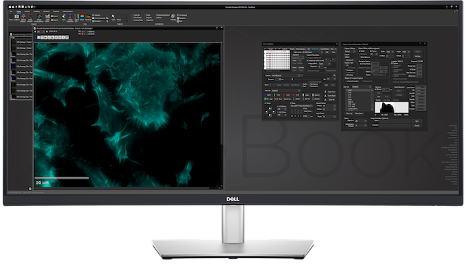
User-Selectable App Appearance

Select a color scheme from dozens of options
Switch on-the-fly from dark to light themes
SlideBook Open File Format

Directory-based open file format for big data and high performance computing applications
Volume Rendering

3D and 4D volume view visualization tools support a user- specified bounding box and a storyboard interface where multiple perspectives can be assembled into a single movie
NVIDIA CUDA GPU Acceleration

GPU acceleration of computationally-intensive operations such as deconvolution
System Capture Consoles
Consoles are a single easy-to-use window featuring all frequent controls and status displays

3D Capture Status
Multiwell and Montage

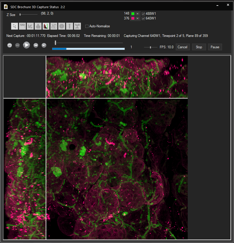
Volumetric projection during 4D capture supported across all instruments
Streamlined multiwell interface
Montaging with a variety of methods


