Nouveau Phasor

All Optical Electrophysiology

All Optical Electrophysiology

Nouveau Phasor is designed for 3D 2-photon stimulation of neurons in vivo. Four distinct modalities enable flexible optogenetics studies involving organisms from C. elegans and Drosophila to zebrafish, mice and larger mammalian species. Nouveau Phasor is able to simultaneously illuminate multiple regions in a 3D volume with the use of SLM-based computer-generated holography.

 
2P Holography 2D-TF 3D-TF 3D-SHOT
Power throughput ••• •• •• ••
3D spatial resolution •• ••• ••
Hand drawn ROIs Yes Yes Yes No
Compatible with any laser Yes Yes Yes Yes
Compatible with fast SLMs Yes Yes Yes Yes
Sample Cell cultures, Plants Brain Slices, Drosophila, Zebrafish Drosophila, Zebrafish, in vivo rodents, Non-human mammals Drosophila, Zebrafish, in vivo rodents, Non-human mammals

Modalities

2P Holography
2P Holography
2D-TF
2D-TF
3D-TF
3D-TF
3D-SHOT
3D-SHOT
2P Holography

2P Holography | Maximum Power

3D Holographic Stimulation

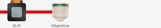
The SLM is exclusively used to modulate the phase of an incoming wavefront from a femtosecond IR laser using the incoming laser’s full power for stimulation patterns.

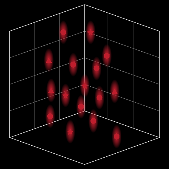
Proprietary software based on iterative Fourier transform algorithms (IFTA) simultaneously generate multiple hand-drawn regions of interest at any position in sample space.

SlideBook’s exclusively licensed libraries generate holographic patterns at multiple Z positions with corrections for power discrepancies inherent to the use of SLMs and each pattern can be assigned a specific power density.

2D-TF

2D-TF | Cell Cultures and Thin Samples

2D Temporal Focusing

A grating is added to the optical path for temporal spreading of the femtosecond beam outside the focal plane. This minimizes the probability of 2-photon absorption above and below the plane of focus.

Temporal Focusing thus offers the possibility to generate axially-confined holographic patterns at the plane of focus.
3D-TF

3D-TF | Flexible Stimulation of Thick Samples

3D Temporal Focusing

A second SLM is added to the optical path.

The first SLM generates the desired patterns, the grating confines the axial extent of the patterns and the second SLM multiplexes the patterns in 3D.

This configuration enables simultaneously-generated axially-confined holographic patterns in 3D.
3D-SHOT

3D-SHOT | Flexible Stimulation of Thick Samples

3D-SHOT

A grating and SLM are used to generate axially-confined Gaussian patterns in sample space. The grating axially confines the Gaussian activation pattern and the SLM multiplexes the pattern at multiple positions in 3D.

A motorized beam expander changes the size of the Gaussian activation pattern.
previous arrow
next arrow

Technology

Computer-Generated Holography

Nouveau Phasor is based on a computer-generated holography (CGH) technique that uses a spatial light modulator (SLM) to modify the phase of an incoming laser beam. It is a highly efficient, phase-only modulation technique that uses all of the power of the incoming laser to generate a holographic pattern at the objective’s focus. Using an iterative Fourier transform, it is possible to apply phase patterns onto the SLM to generate any hand-drawn or computationally outlined pattern in 3D.

Scanless Photostimulation

Scanning photostimulation techniques are generally limited to slower opsins and studies at non-physiological rates. Scanless techniques like holography enable photostimulation across all opsin channels simultaneously so that the cell response speed is only dependent on the cell and the opsin kinetics. As a result Nouveau Phasor enables the use of fast opsins (e.g., Chronos, ChETA, vf-Chrimson, ChroME) allowing for studies at physiological scale with millisecond temporal resolution and sub-millisecond jitter.

Scanning photostimulation techniques are generally limited to slower opsins and studies at non-physiological rates. Scanless techniques like holography enable photostimulation across all opsin channels simultaneously so that the cell response speed is only dependent on the cell and the opsin kinetics. As a result Nouveau Phasor enables the use of fast opsins (e.g., Chronos, ChETA, vf-Chrimson, ChroME) allowing for studies at physiological scale with millisecond temporal resolution and sub-millisecond jitter.

SlideBook Software for Acquisition and Analysis

SlideBook software supports research microscopy through the entire experimental process. By managing everything from instrument control to image processing and data analysis, SlideBook allows scientists to focus on investigation rather than instrumentation. SlideBook controls hundreds of instruments in and around the microscope from dozens of manufacturers enabling researchers to integrate their preferred components and upgrade to the latest devices once available.

Slide

User-Selectable App Appearance

Select a color scheme from dozens of options
Switch on-the-fly from dark to light themes

SlideBook Open File Format

Directory-based open file format for big data and high performance computing applications

Power Calibration

NVIDIA CUDA GPU Acceleration

Uniform distribution of power density in 3D
Geometry calibration of Nouveau Phasor to most research-grade microscope frames

Fast hologram computation for physiological timescales

Stimulation Region of Interests

Hand-drawn or computed stimulation regions at any position in 3D with user-defined power density value

Phasor Streaming

High temporal resolution imaging and simulation sequence recordings

MATLAB Interface

Multiphoton Capture Console

Imaging pipeline for user-defined experimental designs

Consoles are a single easy-to-use window featuring all frequent controls and status displays. The VIVO Multiphoton scanning console also features an intuitive tool for adjusting laser power delivery at different depths with dynamic signal feedback.

Capabilities

Capture

Control hundreds of devices including microscopes, stages, lasers, wheels, piezos, scanners, shutters and much more.

View

Visualize data through any numbers of portals, from single images to z-stacks, time lapse, color channels and 4D views.

Analyze

Analyze images and extract statistical data via a wide variety of algorithms while maintaining original data integrity.

Scripting

Macro scripting for capture and analysis enhances the flexibility and power available to users.

Communicate

Present and export data easily as 16-bit TIFFs, 3D movies, graphs or spreadsheets. Data is directly portable to MATLAB and Excel and adheres to Open Microscopy Environment (OME) standards.

Partners

MATLAB

Through hierarchical and conditional capture, user-supplied MATLAB programs can control experimental workflows.

Microvolution

Microvolution software delivers nearly instantaneous deconvolution by combining intelligent software programming with the power of a GPU.

Aivia

Aivia is an innovative and complete 2D-to-5D image visualization, analysis and interpretation platform with artificial intelligence-guided image analysis.

Dell

The latest high-power computer workstations control all microscope hardware and enable high-speed processing, segmentation and volume rendering of terabyte (TB) datasets.

Powerful Computer Workstation

3i provides high-power computer workstations to control all microscope hardware necessary for acquisition as well as enable processing, segmentation and volume rendering of terabyte (TB) datasets without additional computer resources.  Solid state drives in RAID configurations provide high-speed storage for capture, while traditional high-capacity hard drives provide longer-term storage of datasets. NVIDIA Quadro GPUs work in parallel with the latest Intel Xeon processors for fast stitching, processing and rendering of captured data.

Petabyte Data Storage

3i offers DDN® unified storage systems to allow direct acquisition and analysis, without time-consuming file transfers, at volumes ranging from 500TB to over 2PB. DDN storage systems are an ideal choice for labs and facilities looking to optimize acquisition workflows and/or incorporate data analysis pipelines.

Application Data

Cell Connectivity

Organotypic neurons expressing GCaMP7s and ChroME. Top: 4 cells are simultaneously stimulated at each event. All 4 cells show an increase in calcium activity. One cell responds to the activation without being stimulated (white arrow). Bottom: Sequential activation of each cell shows specific connectivity between cell 4 and the cell shown with the white arrow.

Awake Animal Behavior

3 minute recording of mouse brain calcium activity in vivo via cranial window expressing ChRmine and GCaMP6m. 20 stimulation events were performed for single-cell activation and pair-of-cells activation. All cells had an increase in calcium activity upon stimulation.

Scientific Spotlight

Locomotion

Intrinsic brainstem circuits comprised of Chx10-expressing neurons contribute to reticulospinal output in mice.

Chopek JW, Zhang Y, Brownstone RM. Intrinsic brainstem circuits comprised of Chx10-expressing neurons contribute to reticulospinal output in mice. J Neurophysiol. 2021 Dec 1;126(6):1978-1990. doi: 10.1152/jn.00322.2021. Epub 2021 Oct 20. PMID: 34669520; PMCID: PMC8715053.

Holographic Optogenetic Activation of Neurons Eliciting Locomotion in Head-Embedded Larval Zebrafish.

Jia X, Wyart C. Holographic Optogenetic Activation of Neurons Eliciting Locomotion in Head-Embedded Larval Zebrafish. Methods Mol Biol. 2024;2707:125-140. doi: 10.1007/978-1-0716-3401-1_8. PMID: 37668909.

Learning & Memory

Recurrent architecture for adaptive regulation of learning in the insect brain.

Eschbach C, Fushiki A, Winding M, Schneider-Mizell CM, Shao M, Arruda R, Eichler K, Valdes-Aleman J, Ohyama T, Thum AS, Gerber B, Fetter RD, Truman JW, Litwin-Kumar A, Cardona A, Zlatic M. Recurrent architecture for adaptive regulation of learning in the insect brain. Nat Neurosci. 2020 Apr;23(4):544-555. doi: 10.1038/s41593-020-0607-9. Epub 2020 Mar 23. PMID: 32203499; PMCID: PMC7145459.

Neuronal Cell Activity

Photoactivatable genetically encoded calcium indicators for targeted neuronal imaging.

Berlin S, Carroll EC, Newman ZL, Okada HO, Quinn CM, Kallman B, Rockwell NC, Martin SS, Lagarias JC, Isacoff EY. Photoactivatable genetically encoded calcium indicators for targeted neuronal imaging. Nat Methods. 2015 Sep;12(9):852-8. doi: 10.1038/nmeth.3480. Epub 2015 Jul 13. PMID: 26167640; PMCID: PMC4597790.

Optical vagus nerve modulation of heart and respiration via heart-injected retrograde AAV.

Fontaine AK, Futia GL, Rajendran PS, Littich SF, Mizoguchi N, Shivkumar K, Ardell JL, Restrepo D, Caldwell JH, Gibson EA, Weir RFF. Optical vagus nerve modulation of heart and respiration via heart-injected retrograde AAV. Sci Rep. 2021 Feb 11;11(1):3664. doi: 10.1038/s41598-021-83280-3. PMID: 33574459; PMCID: PMC7878800.

A global timing mechanism regulates cell-type-specific wiring programmes.

Jain S, Lin Y, Kurmangaliyev YZ, Valdes-Aleman J, LoCascio SA, Mirshahidi P, Parrington B, Zipursky SL. A global timing mechanism regulates cell-type-specific wiring programmes. Nature. 2022 Mar;603(7899):112-118. doi: 10.1038/s41586-022-04418-5. Epub 2022 Feb 23. PMID: 35197627.

Regulation of subcellular dendritic synapse specificity by axon guidance cues.

Sales EC, Heckman EL, Warren TL, Doe CQ. Regulation of subcellular dendritic synapse specificity by axon guidance cues. Elife. 2019 Apr 23;8:e43478. doi: 10.7554/eLife.43478. PMID: 31012844; PMCID: PMC6499537.

Comparative Connectomics Reveals How Partner Identity, Location, and Activity Specify Synaptic Connectivity in Drosophila.

Valdes-Aleman J, Fetter RD, Sales EC, Heckman EL, Venkatasubramanian L, Doe CQ, Landgraf M, Cardona A, Zlatic M. Comparative Connectomics Reveals How Partner Identity, Location, and Activity Specify Synaptic Connectivity in Drosophila. Neuron. 2021 Jan 6;109(1):105-122.e7. doi: 10.1016/j.neuron.2020.10.004. Epub 2020 Oct 28. PMID: 33120017; PMCID: PMC7837116.

Two-photon brightness of azobenzene photoswitches designed for glutamate receptor optogenetics.

Carroll EC, Berlin S, Levitz J, Kienzler MA, Yuan Z, Madsen D, Larsen DS, Isacoff EY. Two-photon brightness of azobenzene photoswitches designed for glutamate receptor optogenetics. Proc Natl Acad Sci U S A. 2015 Feb 17;112(7):E776-85. doi: 10.1073/pnas.1416942112. Epub 2015 Feb 4. PMID: 25653339; PMCID: PMC4343171.

In vivo holographic photo-stimulation and two photon GCaMP6 imaging of vagus nerve axons using a GRIN lens integrated nerve cuff.

Futia GL , Fontaine AK, Littich S, McCullough S, Restrepo D, Weir RF, Caldwell JH, Gibson AH. In vivo holographic photo-stimulation and two photon GCaMP6 imaging of vagus nerve axons using a GRIN lens integrated nerve cuff. Proc. SPIE. 2019 Feb 22;Optogenetics and Optical Manipulation 2019, 108660K. doi: 10.1117/12.2521830.

A reorganized GABAergic circuit in a model of epilepsy: evidence from optogenetic labeling and stimulation of somatostatin interneurons.

Peng Z, Zhang N, Wei W, Huang CS, Cetina Y, Otis TS, Houser CR. A reorganized GABAergic circuit in a model of epilepsy: evidence from optogenetic labeling and stimulation of somatostatin interneurons. J Neurosci. 2013 Sep 4;33(36):14392-405. doi: 10.1523/JNEUROSCI.2045-13.2013. PMID: 24005292; PMCID: PMC3761049.

Imaging membrane potential changes from dendritic spines using computer-generated holography.

Tanese D, Weng JY, Zampini V, De Sars V, Canepari M, Rozsa B, Emiliani V, Zecevic D. Imaging membrane potential changes from dendritic spines using computer-generated holography. Neurophotonics. 2017 Jul;4(3):031211. doi: 10.1117/1.NPh.4.3.031211. Epub 2017 May 12. PMID: 28523281; PMCID: PMC5428833.

Additional Resources

Find out more about Nouveau Phasor


    SALES

    Email: sales@intelligent-imaging.com

    Phone: +1 (303)-607-9429 x1

    SUPPORT

    Email: support@intelligent-imaging.com

    Phone: +1 (303)-607-9429 x2